The Core Equation Of Neuroscience
Based on Artem Kirsanov's video on YouTube. If you like this content, support the original creators by watching, liking and subscribing to their content.
Membrane voltage is the primary state variable because it reflects stored electrical energy across the membrane and changes as ionic currents redistribute charge.
Briefing
Neuronal firing can be reduced to a set of coupled differential equations that track how membrane voltage changes as voltage-gated ion channels open and close. The core insight behind the Hodgkin–Huxley framework is that the electrical signal in a neuron is not just a property of “neurons” in general, but of a specific biophysical state variable—membrane potential—whose time evolution is driven by ionic currents. That matters because it turns the messy chemistry of living tissue into a predictive dynamical system: given channel properties and initial conditions, the model can reproduce action potentials and their timing.
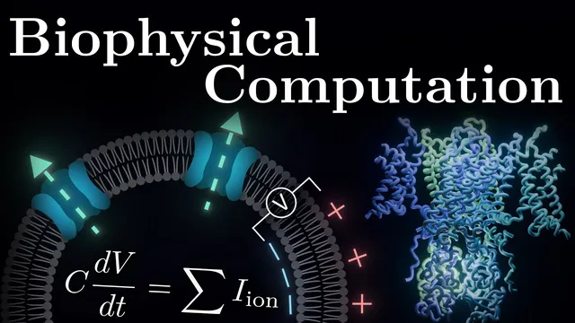
The starting point is a simplified electrical circuit view of a cell membrane. The inside and outside of the membrane hold different ion compositions, leaving more negative charge inside than outside. This charge imbalance creates membrane voltage, which acts like stored electrical potential energy. Because the membrane behaves like a leaky capacitor, voltage changes over time according to how charge redistributes. Real membranes are not perfect capacitors: specialized protein channels allow ions to cross, and those ion flows act like currents. The model links the rate of change of membrane voltage directly to the sum of ionic currents through the membrane.
To make those currents computable, the framework treats each ion’s flow as driven by two competing forces: diffusion from high to low concentration and the electrical attraction or repulsion created by charge separation. For each ion, there is an equilibrium potential where the electrical force balances diffusion, producing zero net current even though ions still move back and forth. When the membrane voltage differs from that equilibrium value, the difference becomes the “driving force,” and the current is proportional to it. The proportionality constant is conductance, which depends on how many channels are open.
Voltage-gated conductance is modeled through gating variables that represent the probability of channel subunits being in permissive states. Hodgkin and Huxley introduce empirical, voltage-dependent opening and closing rate constants (α and β) that govern how these probabilities evolve over time. A key structural result emerges from fitting: potassium channel conductance scales with the fourth power of a single gate probability (n⁴), implying four independent permissive subunits. Later structural biology confirmed that potassium channels indeed contain four identical subunits working together.
Sodium currents follow the same formal logic but with different gate architecture. Sodium conductance depends on multiple gating variables: it scales with M³H, where M activates with depolarization and H represents inactivation that closes under sustained depolarization. With these ingredients, the full Hodgkin–Huxley model becomes a system of four coupled differential equations—one for membrane voltage and three for gating variables—capable of simulating the rise and fall of action potentials.
The model can also be extended beyond a single “point” neuron by dividing complex dendritic and axonal structures into spatial segments and coupling them through additional current-flow terms. That increases realism but raises mathematical complexity. The tradeoff is central: the most complete description is high-dimensional and hard to visualize directly, motivating later reductions to fewer variables that preserve excitability while enabling geometric intuition about how neurons transition between resting and firing states.
Cornell Notes
The Hodgkin–Huxley model turns neuronal firing into a dynamical system by tracking membrane voltage and voltage-dependent ion-channel gating. Membrane voltage changes because ionic currents flow through channels whose conductance depends on how likely channel gates are to be permissive. For each ion, current is proportional to the driving force (membrane voltage minus the ion’s equilibrium potential), with diffusion and electrical forces determining that equilibrium. Potassium conductance scales as n⁴, reflecting four permissive subunits, while sodium conductance scales as M³H, combining activation (M) and inactivation (H). Together these yield four coupled differential equations that can reproduce action potentials and, with spatial extensions, account for complex neuron morphology.
Why is membrane voltage the central state variable in the Hodgkin–Huxley framework?
How do diffusion and electrical forces combine to determine ionic current direction?
What does “driving force” mean mathematically in this model?
Why does potassium conductance scale as n⁴, and what is n?
How do sodium activation and inactivation differ in the Hodgkin–Huxley equations?
What changes when the neuron is modeled as spatially extended rather than a single point?
Review Questions
- How does the equilibrium potential determine the sign of net ionic current for a given ion?
- What assumptions about gate independence lead to conductance scaling like n⁴ for potassium?
- Why does adding spatial compartments make the Hodgkin–Huxley model harder to visualize or solve analytically?
Key Points
- 1
Membrane voltage is the primary state variable because it reflects stored electrical energy across the membrane and changes as ionic currents redistribute charge.
- 2
Ionic current is proportional to the driving force (membrane voltage minus the ion’s equilibrium potential) multiplied by conductance.
- 3
Diffusion and electrical forces compete; their balance at equilibrium potential produces zero net current despite ongoing microscopic ion motion.
- 4
Voltage-dependent conductance is modeled through gating probabilities governed by voltage-dependent opening and closing rate constants (α and β).
- 5
Potassium channel conductance scales with n⁴ because four permissive subunits must align for the channel to conduct.
- 6
Sodium channel conductance scales with M³H, combining voltage-activated opening (M) with voltage- and time-dependent inactivation (H).
- 7
Extending the model to dendrites and axons requires spatial coupling between compartments, turning the system into many coupled differential equations.